Anterior cervical discectomy and fusion
Description
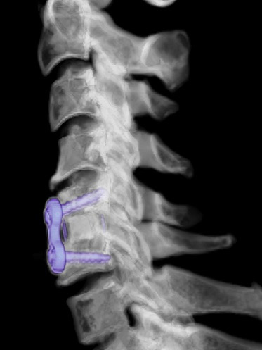
- An anterior cervical discectomy and
 fusion (ACDF) is a surgical procedure performed on the cervical spine (neck) from the front. This approach has several benefits such as better access to the spine and less postoperative pain. Dr. Thapar makes an incision in the front of the neck (usually off to the right) that is generally one to two inches in length. The contents of the neck are gently retracted to the side to expose the cervical spine. The disc material is removed from between the vertebrae using an operating microscope. A synthetic spacer is then sized based on each individual patient’s anatomy. The spacer is filled with bone (both from the patient and from the bone bank) to help facilitate fusion. A plate is positioned over the spacer and screws are inserted to hold the plate and spacer in place.
- X-rays are taken throughout the surgery to determine the correct level and ensure proper size and placement of the spacer, plate, and screws.
Indications
- Herniated disc
- Painful disc degeneration
- Radicular syndrome
- Spondylosis
- Stenosis
- Myelopathy
What to Expect
What to expect before surgery
- You will be fitted for a cervical collar that you will wear after surgery.
- You may need to have a CT scan of the cervical spine if not already done.
- You will need pre-operative clearance from your primary care provider. They will instruct you on any medications that should be stopped prior to surgery such as aspirin and anti-inflammatory medications (Motrin, Advil, ibuprofen, naproxen).
- Do not have anything to eat or drink for at least six hours prior to your scheduled surgery.
- On the day of surgery, an anesthesiologist will meet with you to discuss general anesthesia.
What to expect during surgery
- The operation will take between three and five hours depending on the number of levels.
- The circulating nurse in the operating room will update your family throughout the procedure.
- A CT-like scan is done in the operating room to ensure optimal position of the spacer, plate, and screws.
What to expect after surgery
- You will be in the recovery room for approximately one hour.
- A member of our team will contact your family while you are in the recovery room.
- Your pain will be managed with a Patient Controlled Analgesic (PCA) pump. This will administer a specific amount of medication to you through your IV but also allows you to self-administer additional pain medication.
- You will have a CT scan the morning after surgery.
- You may have to wear a cervical collar after surgery for a variable of time.
- The hospital stay is generally an overnight stay. The majority of patients go home the following day.
- You may have some difficulty swallowing after surgery, this will improve over time.
- You will be given discharge instructions on specific restrictions.
- Recovery after this surgery varies greatly among patients and is dependent on the extent of surgery as well as the age and health of the individual patient. Return to work also varies and is related to overall health and the type of work you do.
POSTERIOR CERVICAL FUSION
Description
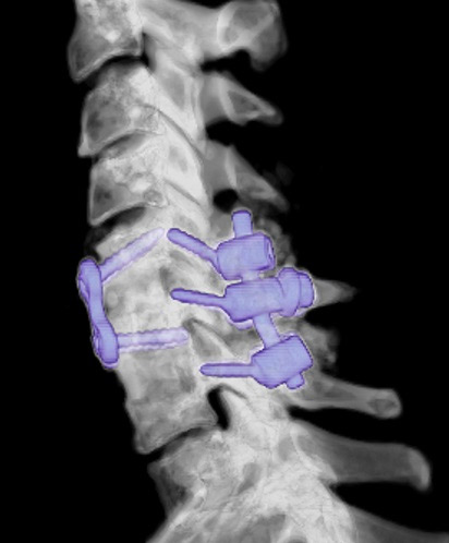
- A posterior cervical fusion is a
 surgical procedure performed on the cervical spine (neck) from the back. Dr. Thapar makes a vertical incision on the back of the neck which is generally three to four inches in length. The muscles of the neck are gently retracted to expose the cervical spine. Screws are placed into the vertebrae to provide stability. A rod is placed on each side to help secure the screws in place. Bone graft is placed along the screws to help facilitate fusion. During the procedure, bone marrow is taken from the iliac crest (hip bone) through a small incision. The bone marrow is used to help facilitate fusion.
- X-rays are taken throughout the surgery to determine the correct levels as well as the size and placement of the screws.
Indications
- Spondylosis
- Spinal stenosis
- Nerve compression
- Fracture of the cervical spine
- Lack of fusion from an anterior approach
What to Expect
What to expect before surgery
- You will be fitted for a cervical collar that you will wear after surgery.
- You may need to have a CT scan of the cervical spine if not already done.
- You will need pre-operative clearance from your primary care provider. They will instruct you on any medications that should be stopped prior to surgery such as aspirin and anti-inflammatory medications.
- Do not have anything to eat or drink for at least six hours prior to your scheduled surgery.
- On the day of surgery, an anesthesiologist will meet with you to discuss general anesthesia.
What to expect during surgery
- The operation will take between three and five hours depending on the number of levels.
- The circulating nurse in the operating room will update your family throughout the procedure.
- A CT-like scan is done in the operating room to ensure optimal position of the spacer, plate, and screws.
What to expect after surgery
- You will be in the recovery room for approximately one hour.
- A member of our team will contact your family while you are in the recovery room.
- Your pain will be managed with a Patient Controlled Analgesic (PCA) pump. This will administer a specific amount of medication to you through your IV but also allows you to self-administer additional pain medication.
- You will have a CT scan the morning after surgery.
- You will may have to wear a cervical collar after surgery for a variable amount of time.
- The hospital stay is generally two to three days.
- You will be given discharge instructions on specific restrictions.
- Recovery after this surgery varies greatly among patients and is dependent on the extent of surgery as well as the age and health of the individual patient. Return to work also varies and is related to overall health and the type of work you do.
Microdiscectomy
Description
- A microdiscectomy is a surgical procedure that is performed when there is a disc herniation present in the lumbar spine that is unresponsive to nonsurgical interventions. A microdiscectomy relieves compression of the spinal nerve by removing the portion of the disc that is herniated or bulging beyond its usual boundaries. Dr. Thapar will make a small vertical incision, generally one to two inches, in the back and retract the muscles to expose the spine. This procedure is done in a minimally invasive fashion. Dr. Thapar will use an operating microscope to identify the area of herniation and remove the disk tissue that is causing the symptoms.
- X-rays are taken throughout the surgery to determine the correct level and the amount of disc tissue that is removed.
Indications
- Herniated or bulging disc in the lumbar spine.
What to Expect
What to expect before surgery
- You will need pre-operative clearance from your primary care provider. They will instruct you on any medications you should stop prior to surgery.
- Do not have anything to eat or drink for at least six hours prior to your scheduled surgery.
- On the day of surgery, an anesthesiologist will meet with you to discuss general anesthesia.
What to expect during surgery
- The operation will take approximately two hours.
- The circulating nurse in the operating room will update your family throughout the procedure.
- The procedure is generally performed through a minimal access approach. This approach allows the surgeon to perform the surgery through a small tube allowing for a smaller incision and less postoperative pain.
What to expect after surgery
- You will be in the recovery room for approximately one hour.
- A member of our team will contact your family while you are in the recovery room.
- Your pain will be managed with a Patient Controlled Analgesic (PCA) pump. This will administer a specific amount of medication to you through your IV but also allows you to self-administer additional pain medication.
- The hospital stay is generally one day.
- You will be given discharge instructions on specific restrictions.
- Recovery after this surgery varies greatly among patients and is dependent on the extent of surgery as well as the age and health of the individual patient. Return to work also varies and is related to overall health and the type of work you do.
laminectomy/decompression
Description
- A decompression is a surgical procedure that is performed to remove compression off the nerve roots or occasionally the spinal cord itself. Dr. Thapar will make a small vertical incision, generally one to two inches, in the back and retract the muscles to expose the spine. An operating microscope to identify the area of compression and remove the bone or other structures that are causing the symptoms.
- X-rays are taken throughout the surgery to determine the correct level and the amount of bone that is removed.
Indications
- Spinal stenosis
- Radicular pain
- Spondylosis
- Neurogenic claudication
What to Expect
What to expect before surgery
- You will need pre-operative clearance from your primary care provider. They will instruct you on any medications you should stop prior to surgery.
- Do not have anything to eat or drink for at least six hours prior to your scheduled surgery.
- On the day of surgery, an anesthesiologist will meet with you to discuss general anesthesia.
What to expect during surgery
- The operation will take approximately two to three hours depending on the number of levels involved.
- The circulating nurse in the operating room will update your family throughout the procedure.
- The procedure is generally performed through a minimal access approach. This approach allows the surgeon to perform the surgery through a small tube allowing for a smaller incision and less postoperative pain.
What to expect after surgery
- You will be in the recovery room for approximately one hour.
- A member of our team will contact your family while you are in the recovery room.
- Your pain will be managed with a Patient Controlled Analgesic (PCA) pump. This will administer a specific amount of medication to you through your IV but also allows you to self-administer additional pain medication.
- The hospital stay is generally one to two days.
- You will be given discharge instructions on specific restrictions.
- Recovery after this surgery varies greatly among patients and is dependent on the extent of surgery as well as the age and health of the individual patient. Return to work also varies and is related to overall health and the type of work you do.
Anterior lumbar interbody fusion
Description
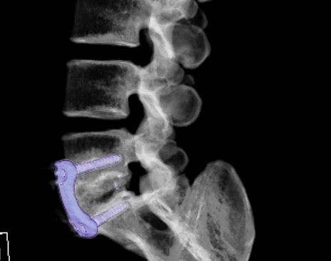
- An anterior lumbar interbody
 fusion (ALIF) is a surgical fusion procedure performed on the lumbar spine (low back) from the front. A vascular surgeon, Dr. Brent Wogahn, performs the exposure of the spine for this type of surgery. The vascular surgeon makes an incision on the lower abdomen. The abdominal contents and major blood vessels are gently positioned off to the side to expose the lumbar spine. Dr. Thapar then removes the disc material between the vertebrae. A spacer is then sized based on each individual patient’s anatomy. The spacer is filled with bone (both from the patient and from the bone bank) and sometimes bone morphogenic protein (BMP) to help facilitate fusion. BMP is a protein that is found naturally in the body and can now by synthetically produced to help promote bone growth. The spacer is secured with either just screws or a plate and screws depending on the level on which the surgery is performed.
- X-rays are taken throughout the surgery to determine the correct level and ensure proper size and placement of the spacer, plate, and screws.
Indications
- Disc degeneration
- Spondylolisthesis
- Scoliosis
What to Expect
What to expect before surgery
- You will be fitted for a lumbar brace that you will wear after surgery.
- You may need to have a CT scan of the lumbar spine if not already done.
- You will need pre-operative clearance from your primary care provider. They will instruct you on any medications that should be stopped prior to surgery such as aspirin and anti-inflammatory medications.
- Do not have anything to eat or drink for at least six hours prior to your scheduled surgery.
- On the day of surgery, an anesthesiologist will meet with you to discuss general anesthesia.
What to expect during surgery
- The operation will take between two to three hours depending on the number of levels.
- The circulating nurse in the operating room will update your family throughout the procedure.
- A CT-like scan is done in the operating room to ensure optimal position of the spacer, plate, and screws.
What to expect after surgery
- You will be in the recovery room for approximately one hour.
- A member of our team will contact your family while you are in the recovery room.
- Your pain will be managed with a Patient Controlled Analgesic (PCA) pump. This will administer a specific amount of medication to you through your IV but also allows you to self-administer additional pain medication.
- You will have a CT scan the morning after surgery.
- You will wear a lumbar brace after surgery for approximately three months. You may take the brace off while in bed.
- A physical therapist will work with you during your hospital stay.
- The hospital stay is generally four to five days.
- You will be given discharge instructions on specific restrictions.
- Recovery after this surgery is generally three months but varies greatly among patients and is dependent on the extent of surgery as well as the age and health of the individual patient. Return to work also varies and is related to overall health and the type of work you do.
Posterior lumbar fusion
Description
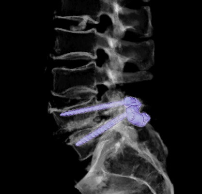
- A posterior lumbar fusion is a
 surgical fusion procedure performed on the lumbar spine (low back) from the back. Dr. Thapar makes a vertical incision on the lower back. The muscles of the back are gently retracted to expose the lumbar spine. Dr. Thapar then places pedicle screws into the vertebrae to provide stability. A rod is placed on each side to help secure the screws in place. Bone graft is placed along the screws to help facilitate fusion.
- A decompression procedure is also commonly performed during this surgery. This involves the surgeon removing compression off the spinal cord or nerve roots with an operating microscope.
- X-rays are taken throughout the surgery to determine the correct level and ensure proper size and placement of the screws.
Indications
- Disc degeneration
- Spondylolisthesis
- Scoliosis
- Fracture of the lumbar spine
What to Expect
What to expect before surgery
- You will be fitted for a lumbar brace that you will wear after surgery.
- You may need to have a CT scan of the lumbar spine if not already done.
- You will need pre-operative clearance from your primary care provider. They will instruct you on any medications that should be stopped prior to surgery such as aspirin and anti-inflammatory medications.
- Do not have anything to eat or drink for at least six hours prior to your scheduled surgery.
- On the day of surgery, an anesthesiologist will meet with you to discuss general anesthesia.
What to expect during surgery
- The operation will take between four and six hours depending on the number of levels.
- The circulating nurse in the operating room will update your family throughout the procedure.
- A CT-like scan is done in the operating room to ensure optimal position of the screws.
What to expect after surgery
- You will be in the recovery room for approximately one hour.
- A member of our teeam will contact your family while you are in the recovery room.
- Your pain will be managed with a Patient Controlled Analgesic (PCA) pump. This will administer a specific amount of medication to you through your IV but also allows you to self-administer additional pain medication.
- You will have a CT scan the morning after surgery.
- You will wear a lumbar brace after surgery for approximately three months. You may take the brace off while in bed.
- The hospital stay is generally four to five days.
- A physical therapist will work with you during your hospital stay.
- You will be given discharge instructions on specific restrictions.
- Recovery after this surgery is generally three months but varies greatly among patients and is dependent on the extent of surgery as well as the age and health of the individual patient. Return to work also varies and is related to overall health and the type of work you do.
Transforaminal lumbar interbody fusion
Description
- A transforaminal lumbar interbody
 fusion (TLIF) is a surgical fusion procedure performed on the lumbar spine (low back) from the back. This surgery allows us to fuse the front and back part of the spine through one surgery and one incision. Dr. Thapar will make a vertical incision on the lower back. The muscles of the back are gently retracted to expose the lumbar spine. The disc material is removed from between the vertebrae and places a graft into the disc space. The graft is filled with bone (both from the patient and from the bone bank) to help facilitate fusion. Dr. Thapar then places pedicle screws into the vertebrae to provide stability. A rod is placed on each side to help secure the screws in place. Bone graft is placed along the screws to help facilitate fusion.
- A decompression procedure is also commonly performed during this surgery. This involves the surgeon removing compression off the spinal cord or nerve roots with an operating microscope.
- X-rays are taken throughout the surgery to determine the correct level and ensure proper size and placement of the graft and screws.
Indications
- Disc degeneration
- Spondylolisthesis
What to Expect
What to expect before surgery
- You will be fitted for a lumbar brace that you will wear after surgery.
- You may need to have a CT scan of the lumbar spine if not already done.
- You will need pre-operative clearance from your primary care provider. They will instruct you on any medications that should be stopped prior to surgery such as aspirin and anti-inflammatory medications.
- Do not have anything to eat or drink for at least six hours prior to your scheduled surgery.
- On the day of surgery, an anesthesiologist will meet with you to discuss general anesthesia.
What to expect during surgery
- The operation will take between four and six hours depending on the number of levels.
- The circulating nurse in the operating room will update your family throughout the procedure.
- A CT-like scan is done in the operating room to ensure optimal position of the screws.
What to expect after surgery
- You will be in the recovery room for approximately one hour.
- A member of our team will contact your family while you are in the recovery room.
- Your pain will be managed with a Patient Controlled Analgesic (PCA) pump. This will administer a specific amount of medication to you through your IV but also allows you to self-administer additional pain medication.
- You will have a CT scan the morning after surgery.
- You will wear a lumbar brace after surgery for approximately three months. You may take the brace off while in bed.
- A physical therapist will work with you during your hospital stay.
- The hospital stay is generally three to four days.
- You will be given discharge instructions on specific restrictions.
- Recovery after this surgery is generally three months but varies greatly among patients and is dependent on the extent of surgery as well as the age and health of the individual patient. Return to work also varies and is related to overall health and the type of work you do.
Extreme lateral interbody fusion
Description
- An extreme lateral interbody
 fusion (XLIF) is a surgical fusion procedure performed on the lumbar spine (low back) from the side. It is a minimally invasive procedure. This surgery allows us to fuse the spine from the side. Dr. Thapar makes an incision on the left side between the ribs and the pelvis. Specific instruments are used to pass through the muscles to the disc space. The disc material is removed from between the vertebrae and a synthetic graft is placed into the disc space. The graft is filled with bone (both from the patient and from the bone bank) and sometimes bone morphogenic protein (BMP) to help facilitate fusion. BMP is a protein that is found naturally in the body that can now be produced synetically that helps promote bone growth. The compression of the spine holds the graft in place.
- A posterior lumbar fusion is commonly performed along with this procedure either on the same or next day.
- X-rays are taken throughout the surgery to determine the correct level and ensure proper size and placement of the graft.
Indications
- Disc degeneration
- Spondylolisthesis
What to Expect
What to expect before surgery
- You will be fitted for a lumbar brace that you will wear after surgery.
- You may need to have a CT scan of the lumbar spine if not already done.
- You will need pre-operative clearance from your primary care provider. They will instruct you on any medications that should be stopped prior to surgery such as aspirin and anti-inflammatory medications.
- Do not have anything to eat or drink for at least six hours prior to your scheduled surgery.
- On the day of surgery, an anesthesiologist will meet with you to discuss general anesthesia.
What to expect during surgery
- The operation will take between two and four hours depending on the number of levels.
- The circulating nurse in the operating room will update your family throughout the procedure.
- You will be positioned on your side.
- A CT-like scan is done in the operating room to ensure optimal position of the screws.
What to expect after surgery
- You will be in the recovery room for approximately one hour.
- A member of our team will contact your family while you are in the recovery room.
- Your pain will be managed with a Patient Controlled Analgesic (PCA) pump. This will administer a specific amount of medication to you through your IV but also allows you to self-administer additional pain medication.
- You will have a CT scan the morning after surgery.
- You will wear a lumbar brace after surgery for approximately three months. You may take the brace off while in bed.
- A physical therapist will work with you during your hospital stay.
- The hospital stay is generally three to four days.
- You will be given discharge instructions on specific restrictions.
- Recovery after this surgery is generally three months but varies greatly among patients and is dependent on the extent of surgery as well as the age and health of the individual patient. Return to work also varies and is related to overall health and the type of work you do.
Carpal tunnel release
Description
- Carpal tunnel release is a surgical procedure performed for carpal tunnel syndrome. Dr. Thapar will make a small incision in the palm of the hand near the wrist to expose the carpal tunnel, which is a passage that holds the median nerve and tendons that flex the fingers. The roof of the transverse carpal ligament is divided to ease the pressure on the nerve.
Indications
What to Expect
What to expect before surgery
- You will need pre-operative clearance from your primary care provider. They will instruct you on any medications you should stop prior to surgery.
- Do not have anything to eat or drink for at least six hours prior to your scheduled surgery.
- On the day of surgery, an anesthesiologist will meet with you to discuss anesthesia. The patient is usually awake during this procedure but given medications through an IV. Local anesthesia is also placed at the surgical site to help relieve pain.
What to expect during surgery
- The operation will take half an hour.
- You will be awake but sedated during the procedure and will likely have no recollection of the procedure.
What to expect after surgery
- You will be in the recovery room for approximately half an hour.
- A member of our team will contact your family while you are in the recovery room.
- Your arm will be wrapped in dressings. Keep the hand clean and dry. Elevate the arm as much as possible.
- You will go home the same day.
- You will be given discharge instructions on specific restrictions.
Ulnar nerve release
Description
- Ulnar nerve release is a surgical procedure performed for ulnar nerve entrapment. Dr. Thapar will make a small incision on the inside of the arm near the elbow. The nerve is moved from its place behind the medical epicondyle to a new place in front of it. This releases the tension of the nerve and prevents it from getting caught on the bony ridge of the elbow.
Indications
What to Expect
What to expect before surgery
- You will need pre-operative clearance from your primary care provider. They will instruct you on any medications you should stop prior to surgery.
- Do not have anything to eat or drink for at least six hours prior to your scheduled surgery.
- On the day of surgery, an anesthesiologist will meet with you to discuss anesthesia.
What to expect during surgery
- The operation will take one hour.
What to expect after surgery
- You will be in the recovery room for approximately one hour.
- A member of our team will contact your family while you are in the recovery room.
- Your arm will be wrapped in dressings. Keep the arm clean and dry. Elevate the arm as much as possible.
- You will go home the same day.
- You will be given discharge instructions on specific restrictions.
 fusion (ACDF) is a surgical procedure performed on the cervical spine (neck) from the front. This approach has several benefits such as better access to the spine and less postoperative pain. Dr. Thapar makes an incision in the front of the neck (usually off to the right) that is generally one to two inches in length. The contents of the neck are gently retracted to the side to expose the cervical spine. The disc material is removed from between the vertebrae using an operating microscope. A synthetic spacer is then sized based on each individual patient’s anatomy. The spacer is filled with bone (both from the patient and from the bone bank) to help facilitate fusion. A plate is positioned over the spacer and screws are inserted to hold the plate and spacer in place.
 surgical procedure performed on the cervical spine (neck) from the back. Dr. Thapar makes a vertical incision on the back of the neck which is generally three to four inches in length. The muscles of the neck are gently retracted to expose the cervical spine. Screws are placed into the vertebrae to provide stability. A rod is placed on each side to help secure the screws in place. Bone graft is placed along the screws to help facilitate fusion. During the procedure, bone marrow is taken from the iliac crest (hip bone) through a small incision. The bone marrow is used to help facilitate fusion.
 fusion (ALIF) is a surgical fusion procedure performed on the lumbar spine (low back) from the front. A vascular surgeon, Dr. Brent Wogahn, performs the exposure of the spine for this type of surgery. The vascular surgeon makes an incision on the lower abdomen. The abdominal contents and major blood vessels are gently positioned off to the side to expose the lumbar spine. Dr. Thapar then removes the disc material between the vertebrae. A spacer is then sized based on each individual patient’s anatomy. The spacer is filled with bone (both from the patient and from the bone bank) and sometimes bone morphogenic protein (BMP) to help facilitate fusion. BMP is a protein that is found naturally in the body and can now by synthetically produced to help promote bone growth. The spacer is secured with either just screws or a plate and screws depending on the level on which the surgery is performed.
 surgical fusion procedure performed on the lumbar spine (low back) from the back. Dr. Thapar makes a vertical incision on the lower back. The muscles of the back are gently retracted to expose the lumbar spine. Dr. Thapar then places pedicle screws into the vertebrae to provide stability. A rod is placed on each side to help secure the screws in place. Bone graft is placed along the screws to help facilitate fusion.
 fusion (TLIF) is a surgical fusion procedure performed on the lumbar spine (low back) from the back. This surgery allows us to fuse the front and back part of the spine through one surgery and one incision. Dr. Thapar will make a vertical incision on the lower back. The muscles of the back are gently retracted to expose the lumbar spine. The disc material is removed from between the vertebrae and places a graft into the disc space. The graft is filled with bone (both from the patient and from the bone bank) to help facilitate fusion. Dr. Thapar then places pedicle screws into the vertebrae to provide stability. A rod is placed on each side to help secure the screws in place. Bone graft is placed along the screws to help facilitate fusion.
 fusion (XLIF) is a surgical fusion procedure performed on the lumbar spine (low back) from the side. It is a minimally invasive procedure. This surgery allows us to fuse the spine from the side. Dr. Thapar makes an incision on the left side between the ribs and the pelvis. Specific instruments are used to pass through the muscles to the disc space. The disc material is removed from between the vertebrae and a synthetic graft is placed into the disc space. The graft is filled with bone (both from the patient and from the bone bank) and sometimes bone morphogenic protein (BMP) to help facilitate fusion. BMP is a protein that is found naturally in the body that can now be produced synetically that helps promote bone growth. The compression of the spine holds the graft in place.